
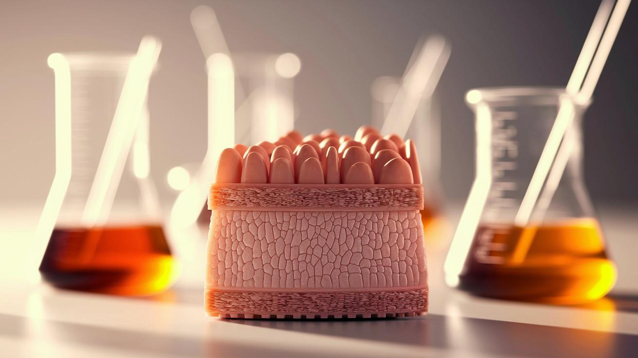
Ever wonder what gives your skin its superpower? Think of it as a cozy pile of layers working together to keep you safe. Picture a simple diagram that shows three layers: the outer epidermis, the middle dermis, and the inner hypodermis. Each layer plays a part like a piece of your very own protective outfit.
Today, we’re chatting about how these layers combine to guard you every day. In plain language, we break down each layer so you can see how your skin acts like nature’s built-in shield. Cool, right?
Visual Guide to Skin Layer Anatomy
Our skin is like a built-in suit of armor, protecting us from bumps, chemicals, heat, cold, UV rays, and germs. It works much like layering your clothes on a chilly day, each layer adds extra safety. Below is a handy diagram that points out the three key layers of your skin:
- Epidermis: the outer shield
- Dermis: offers support and helps you feel touch and temperature
- Hypodermis: keeps you warm with its cushioning and insulation
Imagine putting together your favorite layered outfit where every piece adds comfort and style, each skin layer does something similar to keep you safe. This simple guide turns complex details into an easy-to-understand snapshot, perfect for students, teachers, or anyone curious about how their skin works.
Detailed Epidermis Layer Diagram and Functions
Our skin's top layer, the epidermis, is like a busy, protective shield that renews itself all the time. Most of it is made of keratinocytes, cells that start off vibrant and active, then gradually change through a process called cornification (where they become protective, flattened cells known as corneocytes). It’s a bit like replacing the batteries in your favorite gadget to keep everything running smoothly every day.
Below is a quick guide to the different layers of the epidermis:
| Layer | Description |
|---|---|
| Stratum Basale | This is the deepest layer, packed with active keratinocytes that are firmly attached to the dermis. Think of it as the strong foundation of your skin. |
| Stratum Spinosum | These polygon-shaped cells are linked by tiny connectors called desmosomes, giving your skin its stretch and strength. |
| Stratum Granulosum | Here, cells gather granules that help start a waterproof barrier, much like a raincoat for your skin. |
| Stratum Lucidum | This thin, clear layer is found in thicker skin areas like your palms and soles, adding an extra touch of protection. |
| Stratum Corneum | The outermost layer, made of dead cells that continuously shed, forming the primary barrier against the world outside. |
Each of these layers works closely with the others to keep your skin resilient and well-protected. They move steadily from the active cell growth in the basale right up to the shedding corneocytes in the corneum, ensuring moisture is locked in and invaders like germs and harsh chemicals are kept at bay. It’s nature’s clever way of looking after you every single day.
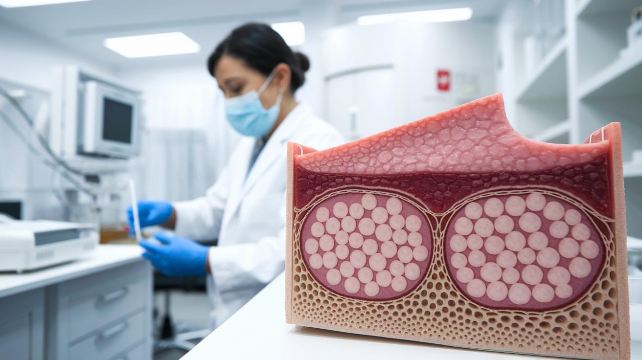
Skin Layers Diagram: Exploring the Dermis Architecture
The dermis is a super important layer of your skin, sitting right under the outer epidermis and being much thicker than its surface counterpart. Its wavy, textured border with the epidermis creates a strong link that not only boosts the exchange of nutrients and moisture but also keeps everything securely in place. This smart design not only holds the layers together but also helps protect your skin from everyday bumps and scrapes. Inside the dermis, you’ll find connective tissues, hair follicles, and oil glands that all add to its unique and functional structure.
Within this layer, a network of fibers provides both firm support and flexibility, making your skin feel resilient and smooth. Tiny blood vessels deliver essential nutrients, while nerve endings take care of sensing touch and temperature, ensuring your body always stays in the loop. Your skin also features different types of sweat glands along with other secretory parts that help keep you cool and maintain hydration. Plus, little lymphatic vessels work quietly in the background to clear away waste. All these elements join forces to keep your skin moist, balanced, and finely tuned to the world around you. This clear diagram of the dermis is a fantastic resource for students, educators, and health professionals who want to understand how our skin’s inner framework contributes to overall wellness.
Hypodermis Layer Diagram: Insulation and Support
Beneath your skin’s outer layers lies the hypodermis, a soft layer mostly filled with fat cells called adipocytes (that’s just a fancy word for little fat stores). These cells gather in clusters that can be thicker in some spots, like on your abdomen and thighs, and thinner in others. Think of this layer as a comfy cushion that not only helps keep your skin in place but also smooths out your natural shape.
This layer acts like a built-in heater on chilly days, trapping warmth to keep you cozy. It also serves as a gentle buffer for your internal organs, absorbing shocks like a natural set of bumpers. Plus, it saves energy for later use, making it a clever helper in your body’s everyday functions.
Variations in Skin Layers Diagram Across Body Regions
Our skin isn’t the same everywhere, it changes in cool, unexpected ways. For example, the skin on your palms and soles, known as glabrous skin, is tougher with a thicker top layer and supportive middle layer. This makes it smooth and hairless, perfect for handling everyday wear and tear. Meanwhile, the rest of your skin, which has more hair follicles and oil glands, is a bit lighter and softer. And then there’s the delicate skin around your eyelids; its outer layer is super thin, almost like a whisper, and has barely any fat underneath.
These differences aren’t random at all, they help your body do exactly what it needs to. The thicker skin on your hands and feet stands up to constant pressure and friction, while the thinner skin in places like your eyelids lets you move easily and gracefully. When you look at a diagram of these skin variations, it’s like seeing a map of how your body is perfectly designed to handle different jobs.
Interactive Skin Layers Diagram Resources for Learning

Dive into skin anatomy with our 3D model that turns learning into a hands-on adventure. You can spin the model, highlight individual layers, and even jot down your own notes, as if you’re exploring a digital lab that lets you zoom in on every tiny detail.
Need something to revisit later? We offer high-quality downloads like a neat PDF diagram and crisp PNG images. Plus, there’s a quiz bank with over 1,800 multiple-choice questions to help you check your knowledge and truly understand how your skin works.
Final Words
In the action, we explored how each skin layer plays a part in shielding and supporting our body. We looked at the epidermis’ quick cell turnover, the dermis’ network of fibers and nerves, and the hypodermis’ cushioning role.
This skin layers diagram helped break down complex ideas into a clear, visual guide that speaks to both style and health. Embrace these insights to feel more confident in everyday choices and enjoy the blend of practicality and chic living.
FAQ
Q: What are the three main layers of skin?
A: The three main layers are the epidermis, dermis, and hypodermis, which work together to protect against injuries, chemicals, and harmful elements.
Q: What has 7 layers of skin?
A: The idea of seven layers usually involves counting the five layers within the epidermis along with the dermis and hypodermis to provide a detailed picture of skin structure.
Q: What are the 5 layers of the epidermis of the skin?
A: These layers include the stratum basale, stratum spinosum, stratum granulosum, stratum lucidum (only in thicker skin), and stratum corneum, all contributing to the skin’s protective barrier.
Q: What layer of skin feels pain?
A: The dermis feels pain because it contains nerve endings that detect sensations like pain, temperature, and pressure.